Familiarize yourself with
the structure of a G protein.
It is composed of 3 subunits
(yellow alpha, purple beta and red gamma)
and is shown here bound with GDP (in CPK colors).
Flip it along the X axis:
Now along the Y axis:
Total Protein Structure
Look at Secondary Structure:
In Jsmol, an
alpha helix is shown
as a magenta spiral, like this:

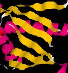
and a
beta pleated sheet is a series of yellow ribbons as shown below
within the context of some alpha heleces:
Focus on the circularized sevenfold propellar shape.
Rotate around the X axis:
Rotate around the Y axis:
What secondary structure is needed to produce this shape?
GTP/GDP Binding Site
GTP in CPK colors
C O
N P
|
GDP is in the Binding Site
Zoom in to see GDP (cpk colors)
bound to the yellow alpha subunit.
Note the two phosphates
Given the quaternary structure shown, why does it make sense that GDP is bound instead of GTP?
Let's focus on the amino acids that bind to GDP
.
You can mouse over each colored amino acid to identify it
(small window appears with three
letter code for amino acids in brackets).
Can you figure out where the third phosphate would be located when GTP is bound to the alpha subunit? Which amino acid(s) would be closest?
Integrating Concepts in Biology
Biology Home Page
© Copyright 2015 Department of Biology, Davidson College, Davidson, NC 28036
Send comments, questions, and suggestions to: macampbell@davidson.edu