Ribozyme vesicles
Goals
- Create the "simplest possible protocell" capable of having a self-replicating informational molecule and a mechanism for spatial localization such as compartmentalization (Chen et al., 2005).
- Use membrane boundary that can grow and divide with being too complex and that can allow passive diffusion of ion and substrates
- Encapsulation of catalytic (self-replicating) RNA molecules within self-replicating membrane vesicles.
Experimental Design
A unique and beneficial aspect of fatty acid vesicles is that they have autocatalytic growth and can repeatedly divide on their own. The first issue addressed is to create membranes that are stable but can allow passive diffusion of ions and substrates in and out of the vesicle. The reason that this aspect of the protocell is so essential is because the formation of RNA catalysts requires the addition of magnesium ions to create the tertiary structure of the ribozyme. To accomplish this goal, researchers observed the effects of magnesium on the stability and permeability of vesicles consisting of fatty acids known as myristoleic acid (MA) and glycerol monomyristoleate (GMM). Thus, they experimented with different ratios of MA to GMM to increase tolerance of Mg2+ in vesicles and allow for passive diffusion.
| MA:GMM ratio | [MgCl2] tolerated, assayed by dye leakage (mM) | [MgCl2] at turbidity change (mM) |
|---|---|---|
| 1:0 | 0.5 | 1 |
| 4:1 | 2 | 3 |
| 2:1 | 4 | 6 |
Table 1. To test the stability of various composititons of MA and GGM, investigators monitored dye retention in the vesicle <1 h after addition of MgCl2. The concentration of MgCl2 that caused leakage to occur is defined as the maximum concentrated tolerated by the vesicle. An additional measure of the maximum concentration of MgCl2 allowed by the vesicle is using the turbidity to access the cloudiness created by individual particles. Table 1 was re-created using data from Chen et al., 2005.
The stability in the presence of Mg2+ was shown to increase as the proportion of GMM increased. However, higher proportions than 2:1 MA to GMM resulted in "the appearance of oil droplets mixed with vesicles" (Chen et al., 2005). Then, researchers were interested in testing the effects of Mg2+ on the permeability of the vesicles. First, they needed to address whether Mg2+ caused permanent permeability in vesicles. Therefore, they measured the percent of dye leakage of vesicles over time. Dye leakage was found to increase over time in a period of one day, showing that permeability of the vesicle exists permanently throughout the experiment (Figure 1A and 1B). Then, researchers tested whether "large-scale destabilization" occurs in vesicles due to Mg2+ by measuring presence of RNA decamers tagged with fluroescent labels (Chen et al. 2005). They would expect if destabilization occurs then the RNA would leak out of the vesicles but instead they found that RNA remained in the vesicles (Figure 1C). However, a mononucleotide (H-UMP) of RNA was found to be permeable in the same conditions (Figure 1D). The paper attributes this difference between the permeability of mononucleotide of RNA and larger RNA molecule to neutralization of negative charges in the RNA and stabilization caused by Mg2+ of the membrane and solute interactions, which would prevent RNA molecules from leaking. Another reason not mentioned in the paper could be that larger RNA molecules may be too large to efficiently diffuse out of the vesicles whereas smaller RNA mononucleotides may be able to pass through the semi-permeable membrane.
http://pubs.acs.org/isubscribe/journals/jacsat/127/i38/figures/ja051784pf00001.gif
Figure 1. (A) Leakage of encapsulated calcein, a fluorescent dye, was measured over time with or without 4 mM MgCl2, represented by the blue and black lines, respectively. (B) Fractions of encapsulated versus free calcein that has leaked out of the vesicle at 22 hr. (C) Leakage of encapsulated RNA decamer is shown by the difference between encapsulated and free RNA using size-exclusion chromatography after 19 hr. The red line represents response to 4 mM Mg2+ versus the control without Mg2+ (black line). (D) Leakage of encapsulated H-UMP vesicles was measured over time in response to MgCl2 (red) versus the control (black) without MgCl2. Image Permission Granted by Jack Szostak.
In addition, investigators used similar processes by using a fluorescent dye sensitive to magnesium known as magfura-2 to verify that these vesicles were indeed permeable to magnesium.
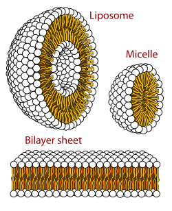
Lastly, researchers attempted to increase vesicle growth by addition of micelles to vesicles. It resulted in a ~50% growth in the surface area of the vesicle. Additionally, dodecane is added as a hydrophobic spacer, resulting in 2:1:0: MA:GMM:dodecane micelles. Thus the overall growth of these micelles to vesicles of the same composition was 40% in one equivalent of micelle.
Results
"Ribozyme Activity in Simple Vesicles"(Chen et al., 2005)
Vesicles of 2:1:0.3 MA:GMM:dodecane were created to encapulate self-cleaving hammerhead ribozymes. This ribozyme (N15min7) is important because it can both cleave and ligate RNA, which will be very important for simple cell-like structures. When Mg2+ is added, the ribozyme cleaves itself into two smaller fragments. The fraction of ribozymes cleaved over time when exposed to 4 mM MgCl2 increased to about 0.66 in unencapsulated vesicles (Figure 2A) and 0.60 in encapsulated vesicles (Figure 2B). The top band on the gel represent the uncleaved ribozymes, while the bottom band represents the cleaved ribozyme, and the lanes correspond with each time point. As the fraction of uncleaved ribozymes decreases, the fraction of cleaved ribozymes increases, which is what we would expect. The vesicles were very stable because even after 15 minutes of exposure to MgCl2, the vesicles remained encapsulated (Figure 2C).
http://pubs.acs.org/isubscribe/journals/jacsat/127/i38/figures/ja051784pf00004.gif
Figure 2. (A and B) The self-cleavage activity of ribozyme N15min7 measured by the fraction cleaved over time. The insets on the graph are phoshorimages of the assay gels. (A) represents unencapsulated ribozymes while (B) represents encapsulated MA:GMM:dodecane ribozymes. (C) Size-exclusion chromatography of MA:GMM:dodecane vesicles of "radiolabeled N15min7 RNA remained encapsulated 15 min after ther addition of MgCl2" (Chen et al., 2005). Image Permission Granted by Jack Szostak.
Conclusions and Further Experiments
Therefore, these researchers sucessfully created vesicles that are permeable to ions and substrates necessary for proper ribozyme function and showed that catalytic ribozyme activity can occur inside these vesicles without any significant loss of functionality. These novel cell-like vesicles open the doors to exploring new ways of engineering and understanding biological systems.