Riboswitches
Riboswitches are small sequences in mRNA that bind small molecules to regulate translation and occasionally transcription. Riboswitches occur naturally in both eukaryotes and prokaryotes. Desai and Gallivan hoped to find new synthetic riboswitches (riboswitches with new ligand specificities) by creating libraries of mutant riboswitches and using genetic selection to pick the functional ones of interest. Desai and Gallivan also employed riboswitches to screen for the presence of specific small molecules. In theory riboswitches are perfect because the number of aptamers already in existence and our capbaility to engineer new aptamers through rational design.
Design
Reviews of previous research showed that the theophylline aptamer worked in riboswitches in wheat germ, a eukaryote, and Bacillus subtilis, a gram positive bacterium. Desai and Gallivan decided to translate the technology to gram negative bacteria. To do this they cloned the theophylline aptamer five base pairs upstream of the RBS for lacZ. The gene is controlled by a weak promoter and a weak RBS allowing for sensitivity to changes in translation because of the presence of theophylline. The construct was then transformed into E. coli. When theophylline is added, translation should be turned on again.
From Concept to Wet Lab
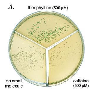
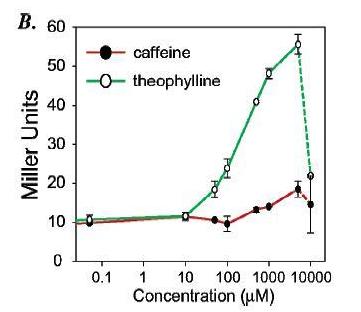
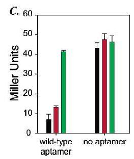
The aptamer for theophylline was inserted in front of the gene for betagalactosidase gene to create an mRNA with a riboswitch. Testing and comparing betagalactosidase activity for cells with the riboswitch in the presence of theophylline, caffeine, and no ligand as well as testing and comparing betagalactosidase activity for cells without the riboswitch under the same conditions demonstrated that the riboswitch does control gene expression in response to theophylline (figure 14). Significant increases in betagalactosidase activity in cells with the riboswitch were only found when theophylline was added. Because betagalactosidase activity in the presence of theophylline, caffeine, or no ligand for cells without a riboswitch did not significantly vary, the increase in betagalactosidase activity in the presence of theophylline for cells with a riboswitch most likely resulted from the theophylline binding to the riboswitch.
Figure 14: a) On the plate are cells with the riboswitch that were grown in three different conditions: theophylline present, caffeine present, and no ligand present. Cells that were grown in the presence of theophylline have a distinct blue/green color compared to the cells grown with caffeine or without ligand. The lack of color in the cells grown with caffeine or without ligand show that the riboswitch prevents translation of the mRNA. The blue color of the cells grown with theophylline indicates that theophylline probably is binding to the riboswitch and allowing for translation. b) The green line represents cells that have had theophylline added to them while the red line represents cells that had caffeine added. Betagalactosidase activity's, as measured by Miller Units, only increasing significantly in the presence of high enough concentrations of theophylline but not caffeine suggests that the riboswitch is highly specific for its ligand. c) When no riboswitch existed in the betagalactosidase transcript, betagalactosidase activity was not significantly different for cells grown in the presence of theophylline, caffeine, or no ligand. When a riboswitch existed in the betagalactosidase transcript, cells grown in the presence of theophylline exhibited significantly greater betagalactosidase activity than cells grown in the presence of caffeine or no ligand. Betagalactosidase activity for cells grown in the presence of caffeine did not vary significantly from activity for cells grown without any ligand present.
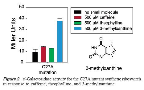
To further support that the theophylline was interacting with the aptamer and not just increasing protein translation through another route. By introducing a single point mutation that in vitro decreases affinity for theophylline and increases affinity for 3-methylxanthine, the lab demonstrated that the translation in the presence of theophylline now was almost the same as translation without any small molecule while translation in the presence of 3-methylxanthine was significantly higher than base-line. These results suggest that the change in translation is controlled by the riboswitch and not some other mechanism.
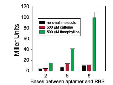
Finally, the lab worked on optimization of the riboswitch by changing the position of the aptamer in relation to the RBS. The riboswitch showed a greater increase in beta-galactosidase when the aptamer was 8 base pairs upstream of the RBS than when the aptamer was either 2 or 5 base pairs upstream. This result could be because of the surrounding bases being purines or it could have something to do with the actual distance.
